Cornea updates 04292022
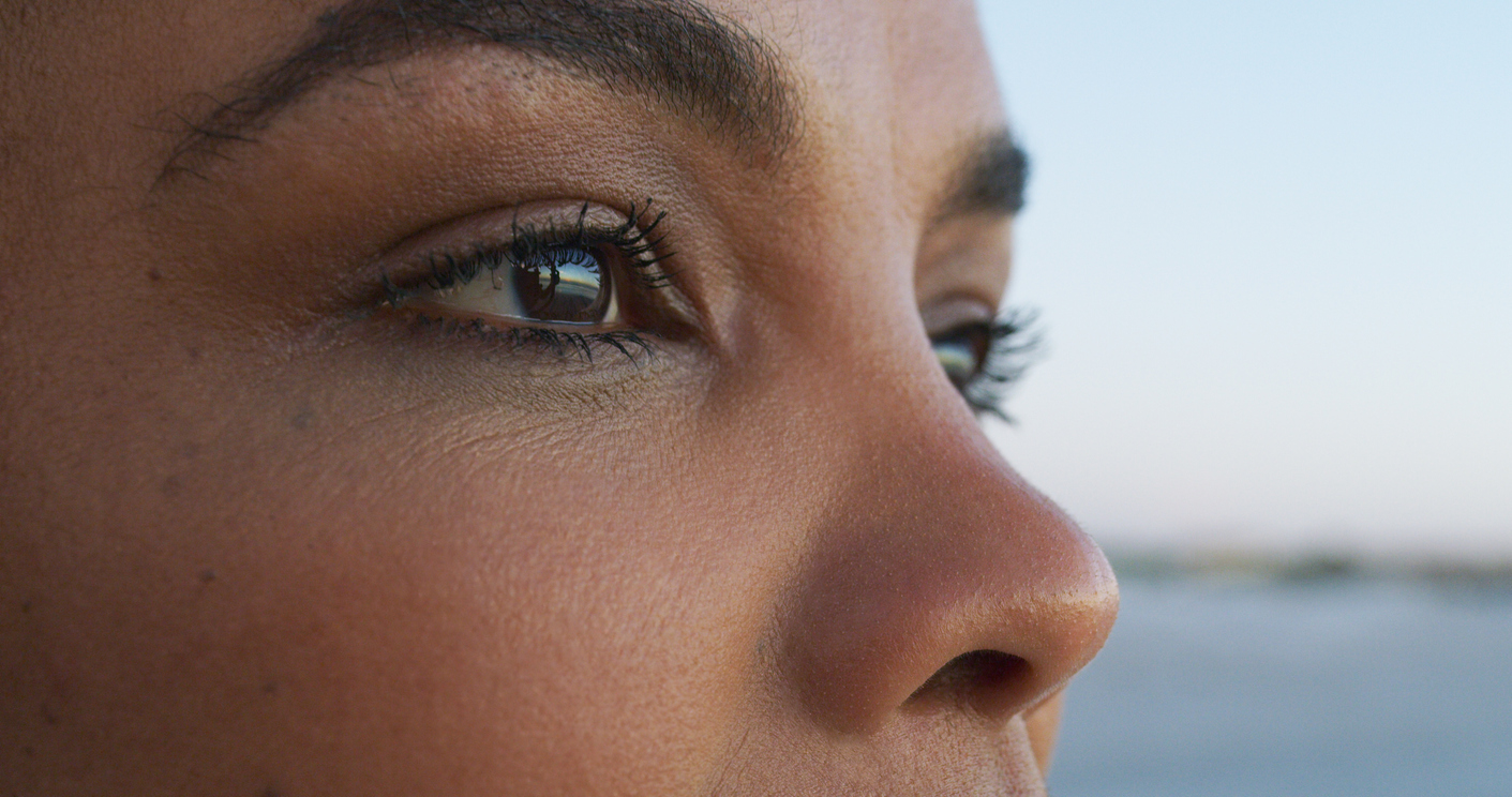
The cornea is the clear front surface of the eye situated directly in front of the iris and pupil. The cornea shields the eye from germs, dust, and other foreign matter and allows light to enter the eye, providing 65% to 75% of the eye’s focusing power. The cornea must be clear, smooth, and healthy for good vision. Most refractive errors (nearsightedness, farsightedness and astigmatism) are a result of less-than-optimal curvature or symmetry of the cornea.
Watch to Learn More About the Cornea
Many of the signs of corneal disorders are the same as other eye conditions:
- Redness
- Blurred/foggy/distorted vision
- Foreign body sensation
- Light sensitivity/glare
- Watery eyes/discharge
- Twitching eyelid
- Pain
Causes of Corneal Disease
- Bacterial, fungal, and viral infections
- Age
- Cataract and intraocular lens (IOL) implant surgery
- Heredity
- Improper contact lens use
- Eye trauma
- Eye diseases
- Dry eye syndrome
- Bell’s palsy and other eyelid disorders
Some common cornea problems include keratoconus, abrasion, infections, scarring, dystrophies, ulcers, pterygium, and others. Your eye doctor will screen for corneal disease and trauma by examining your eyes with magnifying instruments.
Pterygium
Pterygium is a growth of fleshy tissue on the eye’s conjunctiva, the clear covering over the white part of the eye, usually on the side of the eye near the nose. If it grows large enough to cover part of the cornea, it can affect your vision.
Corneal Abrasion
A corneal abrasion is a scratch on the surface of the cornea. Corneal abrasions can be caused by foreign objects as small as a mote of dust. Most corneal abrasions heal completely, with no permanent vision loss, although some deeper abrasions can leave scars and effect vision.
Corneal Erosion
Corneal erosion occurs when the layer of cells on the surface of the cornea, known as the epithelium, detaches from the tissue below. The most common symptom is pain, particularly in the morning. The eye dries while you sleep and the lid can stick to the epithelium, tearing it off when the lid opens. Erosion is more prevalent in people with a history of eye injury or corneal disease, have had an eye ulcer, or wear improperly fitted contact lenses.
Corneal Laceration
A corneal laceration is a cut on the cornea, usually caused by an object striking the eye. If the corneal laceration is deep enough it can cut completely through the cornea and tear the eyeball itself. A corneal laceration is a very serious injury and requires immediate medical attention to avoid severe vision loss. Do not remove the object, rinse the eye with water, or apply pressure to the eye. Do not take aspirin, ibuprofen, or other anti-inflammatory drugs, as they can thin the blood, which can increase bleeding.
Corneal Ulcer
A corneal ulcer (also known as keratitis) is an open sore on the cornea. A corneal ulcer usually results from an eye infection, but severe dry eye or other eye disorders can also cause it. Corneal ulcers can badly and permanently damage your vision and even cause blindness if untreated. If a significant scar remains after the ulcer is healed or an ulcer cannot be treated with medication, a corneal transplant may be needed to improve or keep your vision.
Fuchs’ Dystrophy
Fuchs’ dystrophy is a condition in which the endothelial cells on the back layer of the cornea die. These cells maintain proper fluid levels in the cornea and keep vision clear by pumping out excess fluid. When the endothelial cells die, fluid builds up and the cornea gets swollen and puffy, causing cloudy or hazy vision. In later stages of the disease, tiny blisters may form in the cornea and break open, causing eye pain.
Keratoconus
Keratoconus is a condition in which the cornea progressively becomes thinner, weaker, and irregular or conical in shape. The irregularly shaped cornea projects a distorted image to the brain, resulting in vision loss. Glasses cannot correct vision in patients with keratoconus. Rigid, gas permeable contact lenses are one highly effective way to correct the distortion caused by the irregular cornea, however, in later stages patients may become unable to wear contact lenses and more invasive treatment is necessary. Keratoconus often starts when people are in their teens to early 20s, with vision symptoms slowly worsening over a period of 10 to 20 years. It may occur in only one eye initially, but commonly affects both eyes, with one being more severely affected than the other.
Treatments
Corneal treatments include medication, lubrication, eyeglasses, specialized contact lenses, surgical, or laser procedures.
Intrastromal Corneal Ring Segments (INTACS)
INTACS are a pair of tiny, crescent-shaped, clear plastic implants that are placed into the cornea to flatten the curvature. INTACS provide vision correction and postpone the need for corneal transplant surgery in contact lens intolerant keratoconus patients with adequate cornea thickness and no scarring.
Corneal Cross-linking (CXL)
Corneal cross-linking is an outpatient procedure designed to treat corneal ectasia following refractive surgery and progressive keratoconus. The procedure uses riboflavin (vitamin B2) drops and exposure to ultraviolet light to strengthen and stabilize the cornea to help prevent progression of the condition.
Corneal Transplants
If your cornea is permanently cloudy or scarred, your ophthalmologist may recommend a corneal transplant. This is when the diseased cornea is replaced with a clear, healthy cornea from a human donor. Depending on the which layers of the cornea are diseased or damaged, a full or partial corneal transplant may be required.
Penetrating Keratoplasty (PK) is a full-thickness, complete replacement of the damaged or diseased cornea with a clear donor cornea. A circular area of the central cornea is removed, and the donor cornea is sutured in place with very fine sutures. It can take six to twelve months after a corneal transplant to achieve the best corrected vision.
Descemet’s Stripping Endothelial Keratoplasty (DSEK) and Descemet’s Membrane Endothelial Keratoplasty (DMEK) are partial corneal transplants performed when the innermost layer of the cornea, the endothelium, has been damaged by surgery or is abnormal from Fuchs’ Dystrophy. This cornea transplant technique heals much faster and stronger because only the damaged cell layer is replaced instead of the entire thickness of the cornea.
The damaged/diseased endothelium is stripped from the undersurface of the cornea and replaced with a thin layer of the innermost layers of a donor cornea. This donor tissue is held in place initially with an air bubble, so no sutures are needed to keep it in place. For the first 24 hours after surgery patients need to lie flat on their back as much as possible to help the graft stay in position.
After the first 48 hours there are minimal restrictions to activities. Once the tissue is firmly adhered to the patient’s cornea, it will begin to pump the excess fluid from the cornea and the cornea will begin to clear, usually within one week. The cornea will be 80% healed within one month and vision can continue to gradually improve for four to six months.
Schedule an appointment
Contact us at (803) 779-3070 to schedule an appointment for an eye exam with one of our American Board of Ophthalmology certified physicians at any of our three conveniently located clinics.


 ANNOUNCING UPDATES TO OUR COVID-19 SAFETY PROTOCOLS